Department of Gastroenterology and Hepatology, Princess Alexandra Hospital, Woolloongabba, Queensland, Australia
Department of Gastroenterology and Hepatology, Princess Alexandra Hospital, Woolloongabba, Queensland, Australia
Splenic injury following endoscopy is an interesting however possibly deadly intricacy. While this has been found to happen all the more often after colonoscopy, splenic injury following endoscopic retrograde cholangiopancreatography (ERCP) remains profoundly exceptional since its originally detailed case in 1989. Without a doubt, there have been just 19 such cases revealed in the English, German, and Spanish writing on the whole throughout the course of recent years. We report on a 59-year-elderly person who fostered a peri-splenic haematoma analyzed on stomach registered tomography the day following ERCP and stenting for Mirizzi condition. The patient was dealt with safely and made a full recuperation. We looked into all instances of post-ERCP splenic wounds answered to date and examine the distributed assessments on the logical component of injury, inclining factors, introducing highlights, examination, and treatment choices. Eventually, patient result depends on clinical doubt of this interesting complexity following ERCP.
Splenic injury following endoscopy is an interesting yet possibly deadly entanglement. While this has been found to happen all the more regularly after colonoscopy, splenic injury following endoscopic retrograde cholangiopancreatography (ERCP) remains profoundly exceptional since its previously revealed case in 1989. For sure, there have been just 19 such cases detailed in the English, German, and Spanish writing all in all throughout the course of recent years. We report on a 59-year-elderly person who fostered a peri-splenic haematoma analyzed on stomach registered tomography the day following ERCP and stenting for Mirizzi disorder. The patient was dealt with safely and made a full recuperation. We audited all instances of post-ERCP splenic wounds answered to date and examine the distributed conclusions on the possible instrument of injury, inclining factors, introducing highlights, examination, and treatment choices. Eventually, patient result depends on clinical doubt of this uncommon inconvenience following ERCP.
Keywords: Endoscopic retrograde cholangiopancreatography, Splenic injury, Haematoma
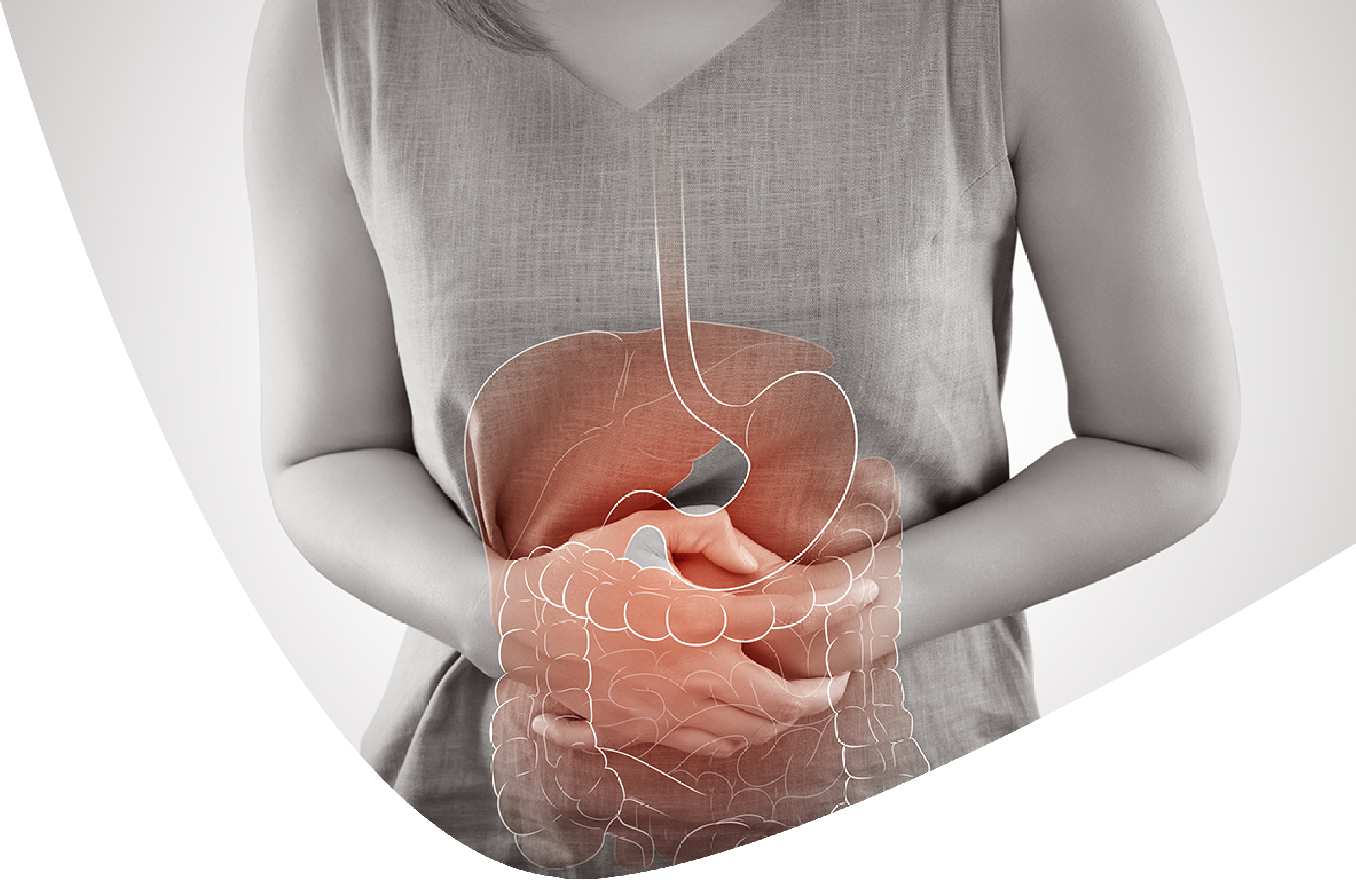
Endoscopic retrograde cholangiopancreatography (ERCP) is a deeply grounded methodology in the administration of pancreaticobiliary conditions. The strategy conveys with it a few all around perceived confusions, as well as other substantially less normal ones, for example, splenic injury, which can be related with critical bleakness or mortality. Starting from the main detailed instance of post-ERCP splenic injury in 1989, there have been a sum of simply 19 cases answered to date in the English, German and Spanish writing [1, 2, 3, 4, 5, 6, 7, 8, 9, 10, 11, 12, 13, 14, 15, 16, 17, 18, 19]. Thus, we report an on a patient peri-splenic haematoma following ERCP and was effectively overseen safely. We likewise audit exhaustively every one of the distributed cases to more readily grasp this intriguing difficulty.
A 59-year-elderly person introduced to the emergency clinic with moderate right upper quadrant stomach torment for 7 days joined by significant jaundice. Before this affirmation, she had introduced to another emergency clinic 7 months already with comparative side effects and was determined to have choledocholithiasis. She went through an ERCP with sphincterotomy and stenting of the normal bile channel however didn't return for an arranged cholecystectomy and expulsion of stent. Her previous clinical history included untreated hepatitis C without cirrhosis, intravenous medication use, liquor misuse and a past laparotomy for ovarian cystectomy. Her standard prescriptions included methadone 80 mg everyday and escitalopram 40 mg day to day. At show, her circulatory strain was 104/70 mm Hg and any remaining crucial signs were inside typical cutoff points.
Actual assessment uncovered a delicate mid-region with delicacy in the right upper quadrant. Her liver capability tests were raised: bilirubin 120 µmol/L (typical, <20 µmol/L), antacid phosphatase 1,490 U/L (ordinary, 30-110 U/L), gamma-glutamyl transferase 850 U/L (ordinary, <38 U/L), alanine transaminase 196 U/L (typical, <34 U/L) and aspartate aminotransferase 145 U/L (ordinary, <31 U/L). Her coagulation studies were all inside typical cutoff points. A liver ultrasound showed cholelithiasis with proof of intrahepatic and proximal extrahepatic bile pipe dilatation. She likewise went through liver attractive reverberation imaging with an attractive reverberation cholangiopancreatography, which uncovered a huge affected stone in the neck of the gallbladder causing impediment of the normal hepatic conduit, only proximal to the recently embedded stent, related with broad pre-stenotic, intra-and extrahepatic ductal dilatation which was with regards to Mirizzi condition. An ERCP was along these lines performed. The stent was taken out with a bushel and the normal bile channel was cannulated effectively through the prior sphincterotomy. The cholangiogram affirmed tight stenosis in the upper third of the primary bile conduit with regards to attractive reverberation cholangiopancreatography discoveries. The injury was stented with a 10 Fr by 12 cm straight plastic stent. The technique was in fact troublesome requiring a supported long extension position to accomplish biliary cannulation and stent situation.
Roughly 4 h after the strategy, routine nursing perceptions recognized recently evolved hypotension with a circulatory strain of 83/50 mm Hg and a pulse of 70 beats/min. The patient was generally asymptomatic, afebrile and griped of no stomach torment. Actual assessment uncovered no stomach delicacy or indications of peritonitis and there was no proof of gastrointestinal draining or unmistakable lack of hydration. Following the organization of 500 mL of intravenous colloid (4% egg whites), her systolic pulse settled at 103 mm Hg which was like that on confirmation. On the following morning, schedule blood tests showed a hemoglobin level of 57 g/L contrasted with 91 g/L before the system (typical, 120-160 g/L). Platelet count and coagulation studies were unaltered and inside ordinary cutoff points. She remained haemodynamically steady and actual assessment was again mediocre, and without proof of gastrointestinal or some other clear wellspring of dying. Considering that the ERCP and stent trade were uninteresting and performed through a prior sphincterotomy, a gastrointestinal draining source was felt to be improbable and a pressing stomach and pelvic processed tomography (CT) examine was coordinated. The CT showed a peri-splenic haematoma and moderate volume of hyperdense stomach free liquid, predictable with a haemoperitoneum. Taking into account she was haemodynamically steady, a choice was made for moderate administration including a blood bonding of 3 units of pressed cells. In the following 48 h, the patient made a uninteresting recuperation and was consequently released from clinic with an arrangement for a possible elective cholecystectomy. She was followed up as a short term 3 weeks after the fact; she was clinically well with typical reach hemoglobin and bilirubin.
Beginning around 1968, when ERCP was first presented by McCune et al. [20], there has been critical progression and expanded utility of this system for analytic and presently for the most part restorative purposes in overseeing pancreaticobiliary conditions. Normal confusions of this strategy as announced by the American Culture for Gastrointestinal Endoscopy (ASGE) in 2012 incorporate pancreatitis (approx. 3.5%), drain (particularly following sphincterotomy; 1.3%), cholangitis (1% or less), cardiopulmonary intricacies (1%), hole (0.1-0.6%) and in general mortality (0.33%) [21]. Splenic-related wounds have forever been viewed as an interesting however perceived confusion of endoscopy, inferable from the nearby physical vicinity of the spleen to the stomach and colon. Other detailed intriguing intricacies of ERCP incorporate liver gash and interruption of the cross over mesocolon with resultant colonic ischaemia [1, 2]. Before 1975, 2 enormous case series including north of 12,000 patients who went through colonoscopy and endoscopy didn't find a solitary instance of splenic injury [3]. Starting around 2014, Weaver et al. [4] revealed that there were north of 60 instances of splenic injury following colonoscopy however just 13 reports of a comparable physical issue after ERCP in the writing.
The principal instance of splenic injury following ERCP was accounted for by Trondsen et al. [5] in 1989. A female patient conceded with intense gallstone pancreatitis was found to have experienced a total separation of the splenic case requiring splenectomy roughly 15 h after ERCP. From that point forward, comparative cases have been accounted for over the accompanying 27 years though of varying patient and procedural attributes. The sorts of splenic wounds that happen are restricted, and the proposed system of injury and inclining factors have had little varying assessment among the creators, as will be examined underneath. At the hour of this report, a sum of 19 instances of ERCP-related splenic injury were seen as in the English, German, and Spanish writing. These distributed cases included 11 female and 8 male patients with a middle age of 57 years (range, 33-82 years).
The specific mechanism(s) of injury to the spleen during ERCP stays unsettled; in any case, unreasonable extension related direct, footing or shear powers show up undoubtedly causative. As the side-review duodenoscope is generally cutting-edge into a circle (long) position to arrive at the second piece of the duodenum, the degree must be twisted to one side and removed to some extent (short position) to bring the ampulla into a palatable situation for cannulation. This move causes beginning "bowing" of the endoscope inside the stomach, logical converting into direct powers towards encompassing organs including the spleen, trailed by twist on the more prominent ebb and flow of the stomach. Nearby viscera that are physically connected, like the spleen, could then be harmed by foothold powers, appearing as splenic capsular tears or vascular separation [6, 7, 8, 9, 10, 11, 12]. A supported long degree position, as for our situation, could in this manner be related with delayed powers.
There are something like [3]inclining factors that have been proposed to prompt splenic injury during ERCP. Diminished development between the spleen and nearby designs, abundance foothold on the gastrosplenic tendon and direct degree incited injury have been guessed [3, 13]. Five cases included patients with either known or recently analyzed (intraoperative) constant pancreatitis, and it has been proposed that calcification and fibrosis of the joining tendons block any compensatory versatility between the encompassing organs [2, 3, 4, 15, 16]. Grips from earlier stomach a medical procedure, as for our situation, were likewise observed to be related with splenic wounds for a similar explanation [2, 12, 13, 17]. As a matter of fact, given the close vicinity between the spleen and tail of pancreas, intense and post-ERCP pancreatitis is progressively perceived as a gamble factor for splenic parenchymal wounds [22]. Actually troublesome cases because of modified life structures or other patient variables might bring about delayed strategy time and more extension control, in this way expanding the gamble of unnecessary ligamentous strain and injury. Stomach expansion itself, according to the creators of no less than 2 cases, may well deliver the most elevated (and briefest) short gastric vessels defenseless against separation injury [2, 14]. On the other hand, direct splenic injury by the duodenoscope is plausible, which frequently brings about injury to the average or hilar surface of the spleen [14].
It is quite important that splenic wounds have happened even in non-in fact troublesome methodology, as featured by Wu and Katon [1] and Chavalitdhamrong et al. [23]. Of the 19 instances of post-ERCP splenic wounds, 10 methods were performed easily, 7 were troublesome, and in 2 it was unreported. Moreover, albeit restorative ERCP generally conveys a higher horribleness and mortality than symptomatic ERCP [1, 21], the thing that matters was not essentially exhibited in our survey. Seven cases just had indicative ERCP performed, while 12 likewise had remedial strategies.
Signs and side effects that ought to make a clinician aware of a potential splenic physical issue are not explicit and cross-over extensively with essentially more normal difficulties of ERCP, for example, pancreatitis, hole and post-sphincterotomy dying. The average show might incorporate stomach torment regardless of indications of peritonitis (frequently left upper quadrant and epigastric), hypotension and additionally tachycardia [1, 2, 3, 4, 5, 6, 7, 8, 9, 10, 11, 12, 13, 14, 15, 16, 17, 18, 19]. Fever was found in just 1 case who addressed 10 days after the fact - in the setting of a formerly known splenic haematoma that turned out to be optionally tainted from a skin source and transformed into a boil [14]. Hemoglobin was in every case altogether diminished (>20 g/L) contrasted with pre-technique levels. It should likewise be underscored that there is an impressively factor time span between creating side effects and file ERCP. While somebody who gripes of intense stomach torment minutes after the methodology would quickly raise doubt of a connected confusion, deferred introductions of as long as 6 days have been accounted for and might be neglected as an irrelevant issue.
Albeit in 5 out of 19 cases, the patient was taken straightforwardly to theater for crisis laparotomy in light areas of strength for of doubt alone, a stomach CT filter is as yet thought to be the trial of decision to research any thought extreme confusion [23, 24]. On the other hand, stomach ultrasound has additionally been utilized in 2 cases which uncovered discoveries that upheld the sign for a medical procedure [5, 13]. Normal pathologic discoveries on CT incorporate moderate to huge volume of free liquid (haemoperitoneum), subcapsular or peri-splenic haematoma, and splenic gash. Frequently the specific finding must be found out intraoperatively, as in cases because of separation of veins and splenic capsular tears.
The board of the patient who has likely experienced a post-ERCP splenic injury doesn't necessarily involve a laparotomy. In this survey, everything except one patient made a full recuperation either following employable or moderate administration. One patient passed on from Pseudomonas pneumonia and resulting multi-organ disappointment after splenectomy [2]. To be sure, the board ought to be custom-made to the patient's clinical state and with respect to their haemodynamic dependability. Injury rules ought to likewise be observed according to any unpolished splenic injury, as the administration is comparative regardless of varying components of injury. Early contribution of the careful group is reasonable for any ERCP-related difficulty, especially for splenic injury. A careful methodology is normally shown in patients who experience haemodynamic split the difference in spite of satisfactory revival, while the people who are clinically steady likely could be noticed and treated moderately by an accomplished clinician. In select cases, a splenorrhaphy as opposed to a splenectomy might be considered at laparotomy for stable patients with a recognizable splenic physical issue that is repairable [3, 10]. All the more as of late, splenic supply route embolisation has arisen as a valuable assistant to non-employable administration and is progressively utilized in injury settings.
The patient for our situation went through an in fact troublesome yet fruitful ERCP with stenting for biliary obstacle, and consequently encountered an episode of hypotension with no stomach torment and a drop in hemoglobin the following day with next to no other concerning highlights. ERCP-related gastrointestinal draining seemed improbable given that biliary cannulation and stenting was performed through a prior sphincterotomy. The CT showed a peri-splenic haematoma with haemoperitoneum, which we suspect was because of splenic capsular injury, reasonable related with powers from a supported long extension position expected to perform ERCP and stent situation effectively. She was dealt with moderately and made a full recuperation.
All in all, splenic injury with splenic haematoma or drain ought to be perceived as an uncommon however clinically critical complexity of ERCP. Clinical doubt ought to be raised in the event that the technique has been troublesome, requiring a supported long place of the duodenoscope. Side effects might incorporate starting asymptomatic post-procedural hypotension, left upper quadrant torment, or potentially particularly decreased hemoglobin with no haematemesis or melaena. There ought to be a low limit for considering CT imaging in this clinical situation. Thought ought to likewise be given for remembering the intriguing gamble of splenic injury for the assent data for ERCP.
Assent for distribution has been acquired from the patient.
The creators pronounce no irreconcilable circumstances.
1. Wu WC, Katon RM. Injury to the liver and spleen after symptomatic ERCP. Gastrointest Endosc. 1993;39:824-827. [PubMed] [Google Scholar]
2. Kingsley DD, Schermer CR, Jamal MM. Interesting inconveniences of endoscopic retrograde cholangiopancreatography: two case reports. JSLS. 2001;5:171-173. [PMC free article] [PubMed] [Google Scholar]
3. Dixon E, Graham JS, Sutherland F, Mitchell PC. Splenic injury following endoscopic retrograde cholangiopancreatography: a case report and survey of the writing. JSLS. 2004;8:275-277. [PMC free article] [PubMed] [Google Scholar]
4. Weaver JL, Jones W, Mill operator KR. Hazardous splenic burst after endoscopic retrograde cholangiopancreatography. Am Surg. 2014;80:E230-E231. [PubMed] [Google Scholar]
5. Trondsen E, Rosseland AR, Moer A, Solheim K. Burst of the spleen following endoscopic retrograde pancreatography (ERCP) Case report. Acta Chir Scand. 1989;155:75-76. [PubMed] [Google Scholar]
6. Ong E, Bohmler U, Wurbs D. Splenic injury as an intricacy of endoscopy: two case reports and a writing survey. Endoscopy. 1991;23:302-304. [PubMed] [Google Scholar]
7. Lewis FW, Moloo N, Stiegmann GV, Goff JS. Splenic injury entangling helpful upper gastrointestinal endoscopy and ERCP. Gastrointest Endosc. 1991;37:632-633. [PubMed] [Google Scholar]
8. Lo AY, Washington M, Fischer MG. Splenic injury following endoscopic retrograde cholangiopancreatography (ERCP) Surg Endosc. 1994;8:692-693. [PubMed] [Google Scholar]
9. Badaoui R, Ouendo M, Delcenserie R, El Kettani C, Radji M, Ossart M. Injury to the liver and spleen after symptomatic ERCP. Can J Anaesth. 2002;49:756-757. [PubMed] [Google Scholar]
10. Zyromski NJ, Camp CM. Splenic injury: an uncommon complexity of endoscopic retrograde cholangiopancreatography. Am Surg. 2004;70:737-739. [PubMed] [Google Scholar]
11. Ahmad S, Bragg D, Zaitoun AM, Lobo DN. A risky circle. Clin Case Rep. 2016;4:535-536. [PMC free article] [PubMed] [Google Scholar]
12. Paredes AH, Williams AM, Vertrees AE, Womeldorph C. Splenic cut following ERCP. Endoscopy. 2013;45:E221-E222. [PubMed] [Google Scholar]
13. Villalobos-Garita A, Alvarado-Salazar M, Rodriguez-Quesada G, Paez-Saenz R, Campos-Goussen C, Barahona-Garcia R, Avalos-Giugliarelli A, Gonzalez-Pacheco O. ERCP and splenic injury. Endoscopia. 2014;26:23-25. [Google Scholar]
14. Furman G, Morgenstern L. Splenic injury and sore muddling endoscopic retrograde cholangiopancreatography. Surg Endosc. 1993;7:343-344. [PubMed] [Google Scholar]
15. Gaffney RR, Jain V, Moyer MT. Splenic injury and ERCP: a potential gamble for patients with cutting edge persistent pancreatitis. Case Rep Gastroenterol. 2012;6:162-165. [PMC free article] [PubMed] [Google Scholar]
16. Cho CL, Yuen KK, Yuen CH, Chong LC, Chu RW. Splenic slash after endoscopic retrograde cholangiopancreatography. Hong Kong Drug J. 2008;14:145-147. [PubMed] [Google Scholar]
17. Grammatopoulos A, Moschou M, Rigopoulou E, Katsoras G. Splenic injury confusing ERCP. Ann Gastroenterol. 2014;27:177-178. [PMC free article] [PubMed] [Google Scholar]
18. Deist TJ, Freytag A. Splenic burst after ERCP. Z Gastroenterol. 2003;41:579-582. [PubMed] [Google Scholar]
19. Cortinas Saenz M, Maldonado Hermosos G, Miota de Llama JI, Moreno Planas JM, City hall leader Lopez JC. Burst of the spleen following endoscopic retrograde cholangiopancreatography. Fire up Esp Enferm Dig. 2010;102:457-458. [PubMed] [Google Scholar]
20. McCune WS, Shorb PE, Moscovitz H. Endoscopic cannulation of the ampulla of Vater: a primer report. Ann Surg. 1968;167:752-756. [PMC free article] [PubMed] [Google Scholar]
21. ASGE Norms of Training Advisory group, et al. Difficulties of ERCP. Gastrointest Endosc. 2012;75:467-473. [PubMed] [Google Scholar]
22. Patil PV, Khalil A, Thaha Mama. Splenic parenchymal difficulties in pancreatitis. J Pancreas. 2011;12:287-291. [PubMed] [Google Scholar]
23. Chavalitdhamrong D, Donepudi S, Pu L, Draganov PV. Remarkable and seldom revealed antagonistic occasions of endoscopic retrograde cholangiopancreatography. Digest Endosc. 2014;26:15-22. [PubMed] [Google Scholar]
24. Pannu HK, Fishman EK. Difficulties of endoscopic retrograde cholangiopancreatography: range of anomalies showed with CT. Radiographics. 2001;21:1441-1453. [PubMed] [Google Scholar]
Richard Lee. Following endoscopic retrograde cholangiopancreatography, there was splenic injury. International Journal of Gastroenterology and Hepatology 2022.