Naval Medical Center Portsmouth, Portsmouth, VA, USA
Naval Medical Center Portsmouth, Portsmouth, VA, USA
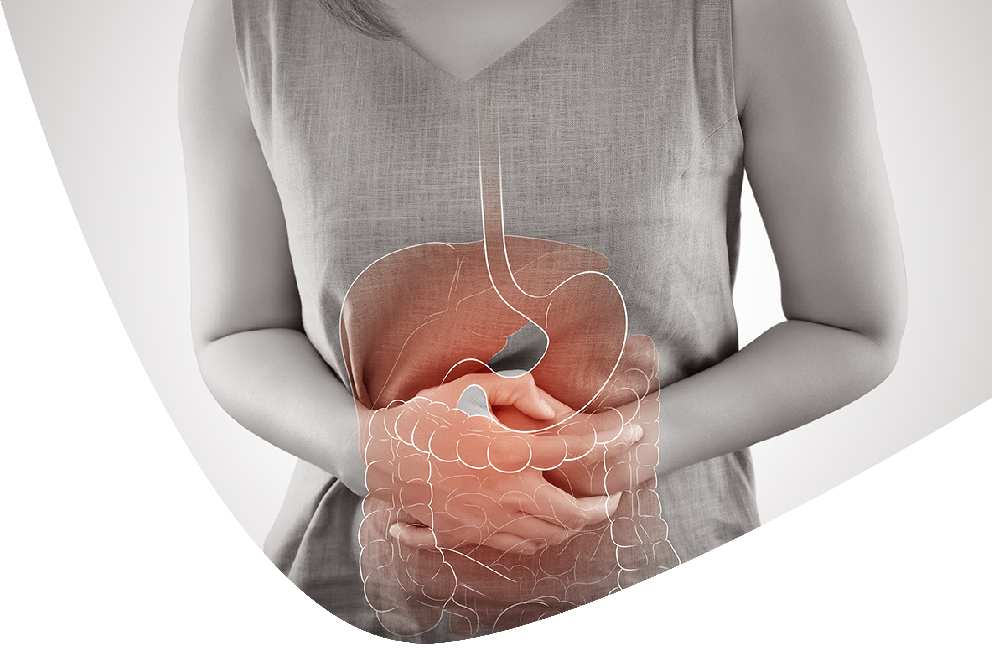
Duodenal variceal draining is a phenomenal intricacy of entrance hypertension that can undoubtedly go unnoticed and arrive at death rates as high as 40%. Cirrhosis is the most widely recognized reason for duodenal varices. Much of the time, duodenal varices happen correspondingly with esophageal varices, further confounding distinguishing proof with beginning endoscopy. Albeit numerous modalities have been investigated concerning the board and treatment draws near, rules still can't seem to be laid out attributable to the rarity where draining happens from ectopic duodenal varices.
Keyphrases: Ectopic varices, Variceal embolization, Upper gastrointestinal dying
Cirrhosis is an irreversible course of liver fibrosis and is liable for huge grimness and mortality around the world. As fibrosis advances, expanded protection from entry venous blood stream creates and gateway hypertension (PHT) can happen. Entanglements of PHT are many, however the most dreaded and intense is a variceal drain. Most of variceal draining happens in the throat or the stomach at a pace of 5-15% yearly [1]. Once in a while, varices are tracked down beyond the gastroesophageal district, and these are alluded to as ectopic varices (ECV). Varices are tracked down in the duodenum, jejunum, ileum, colon, peristomal, rectum, and numerous different areas. Duodenal varices are the most well-known little gut varices, and cirrhosis is the most widely recognized cause. As per one review done by Saad et al. [2] 40% of patients with PHT going through angiography were recognized to have duodenal varices. Discharge from ectopic duodenal varices can be challenging to analyze, frequently requiring rehash endoscopies and imaging studies. Concerning treatment, there are a few modalities using endoscopic procedures that incorporate endoscopic band ligation, sclerotherapy and cutting. Higher hepatic venous tension inclinations are more prescient of treatment disappointment, so, all in all interventional radiologic systems might be expected for control of dying. Medical procedure is thought of if endoscopic and radiologic strategies have fizzled. Forecasts differ by site, and a lot of what it is accounted for in the writing depends on case reports and series. We present an instance of enormous duodenal variceal drain that eventually required a transesophageal intrahepatic portosystemic shunt (TIPS) with curl embolization for control of dying.
A 56-year-old male with decompensated alcoholic cirrhosis Model for End-stage Liver Infection (Merge) 12 at show, was moved to our emergency clinic for the executives of progressing hematochezia notwithstanding getting esophageal variceal ligation (EVL) with 3 groups set for high-risk blemish varices. The patient had an ordinary colonoscopy. The introducing hemoglobin (Hgb) from the moving office was 6.4 g/dL with an ascent to 6.7 g/dL after 2 units of pressed red platelets. On appearance, rehash esophagogastroduodenoscopy (EGD) uncovered dislodgment of the 3 esophageal groups with hidden ulcerations and platelet plugs as well as segments of huge esophageal varices. Also, there was new blood found in the stomach. Five groups were redeployed in the distal throat without any proof of dynamic draining at finishing. The patient was bonded to Hgb 7.3 g/dL yet had rehash hematochezia and resulting decrease in Hgb to 6.2 g/dL the next morning. Interventional radiology was counseled for TIPS arrangement optional to worries for bombed EVL with rebleeding. Rehash new EGD uncovered 3 of the recently positioned groups were unstuck with no dynamic dying. Two medium varices were found in the lower third of the throat that were grouped and effectively collapsed. No draining was imagined in the gastric cardia or fundus. Sketchy friable mucosa was noted in the proximal duodenum. The patient kept on requiring blood bondings and pressor help. A difference upgraded CT examine was performed for TIPS careful preparation, as no earlier cross-sectional imaging was accessible. The CT check uncovered an especially cirrhotic liver, ascites, and beforehand undiscovered numerous unmistakable varices including the third piece of the duodenum . The patient was moved from the CT scanner to the Interventional Radiology Suite and a developing TIPS was effectively performed. During the TIPS system, entrance venography exhibited a somewhat unmistakable coronary vein without clear esophageal varices. There was critical hepatofugal stream in the entryway vein. Moreover, there were conspicuous duodenal varices noted stretching out from the proximal predominant mesenteric vein with shunting of stream towards the left renal vein which compared to the discoveries seen on CT, and logical the wellspring of the patient's kept draining rather than the esophageal varices seen on endoscopy. When the TIPS shunt was set, there was critical decrease in the portosystemic slope from 25-30 to 3-5 mm Hg with a perceptible diminishing in coronary vein widening and return of hepatopetal stream of gateway blood towards the liver. Given the undeniable gateway framework decompression, further mediations were conceded.
On postoperative day 1, status-post TIPS arrangement, the patient balanced out however had intermittent hematochezia with a Hgb nadir of 5.0 g/dL requiring factor VII for inversion of progressing coagulopathy and amendment of INR from 2.5 to 0.9. A push enteroscopy was performed. The earlier groups were unblemished, there was no proof of draining or high-risk blemish, the gastric fundus and cardia were again envisioned, and again there was no proof of varices, dying, or high-risk blemish. Given the discoveries on CT and on angiography, the third piece of the duodenum was assessed showing a huge varix with high-risk blemish of draining seen as proven by discouraged punctate white spot inside an area of shallow erythema.
Attributable to the size of the varix and the intermittent hematochezia, endoscopic treatment was conceded, and the patient went through effective endovascular curl embolization of the duodenal varices . There could have been no further draining postoperatively. He created gentle grade 1 hepatic encephalopathy that answered rifaximin and his Merge score topped at 23. The patient was released home in a steady condition on postoperative day 6 status-post TIPS. He at last required 27 units of stuffed red platelet bondings. The patient was seen most as of late at his 3-month postoperative encounter with a critical improvement in the Merge score, presently at 15. He has been off his rifaximin for a month with next to no episodes of encephalopathy, doesn't have ascites, and has gotten back to work and is completely utilitarian.
ECV are intricate compressed portosystemic venous guarantees happening inside the midsection barring the gastroesophageal locale and record for up to 5% of all variceal dying. Drain of these varices can be huge with a death rate as high as 40% [2, 3]. Ectopic duodenal varices, for example, found for our situation, represent 17% of ECV and 25-33% of ectopic drains. Cirrhosis is the most widely recognized reason for duodenal varices, representing almost 30% of cases [2]. It is likewise imagined that duodenal varices might create after EVL because of unconstrained portosystemic shunting to substitute destinations. Most of varices are found in the duodenal bulb and the plunging part of the duodenum [3]. The recurrence of draining and event decline in the more distal segments of the duodenum in light of the fact that duodenal varices are found somewhere down in the serosal layer though esophageal varices are situated in the submucosal layer. ECV can give hematemesis, hematochezia and as dark drains relying upon the area of the varices. Analysis can be testing, and up to 69% of individuals with duodenal varices might have coinciding esophageal varices. Upwards of 5 rehashed EGDs have been expected to make the determination [4].
Ideal administration isn't deeply grounded, and there are no treatment rules for ECV. Ectopic variceal hemorrhages ought to be overseen similarly as gastroesophageal hemorrhages regarding liquid revival, anti-microbials, and octreotide with vasopressors if necessary [1]. Endoscopic sclerotherapy has been effectively utilized in the past with great results, albeit this conveys a gamble of hole. EVL has likewise demonstrated effective in halting dying; notwithstanding, this approach is restricted in varices bigger than 15 mm, similarly as with this patient, in light of the fact that getting a decent view is periodically troublesome, and segregating the taking care of vessels can be hazardous. Endoscopic hemoclips can be sent and control draining by giving direct mechanical tension in the event that the afferent and efferent vessels can be secluded. Entanglements incorporate potentiating draining and conceivable hole whenever set erroneously. TIPS is viable in controlling draining in the intense setting yet has no mortality benefit over endoscopic treatments. A little report has exhibited that patients with hepatic venous strain slopes more noteworthy than 20 mm Hg have a critical improvement in endurance when performed inside 24 h of an intense variceal drain yet further examinations to approve this are required [1]. TIPS alone has all the earmarks of finding success as rescue treatment and diminishes the requirement for rehashed strategies, and is demonstrated powerful in forestalling repetitive esophageal variceal rebleeding [4]. TIPS is additionally suggested by the American Relationship for the Investigation of Liver Illnesses for the anticipation of rebleeding in ECV. There is anyway a pattern towards expanded rebleeding-related mortality found in the TIPS alone gathering. When joined with embolization, there is better result over the TIPS alone gathering in anticipation of rebleeding in ECV [2, 5, 6]. Sadly, post-TIPS hepatic encephalopathy and shunt stenosis are known confusions, and results are more regrettable in patients with further developed Youngster Pugh scores [4].
All in all, early angiographic ID and mediation can't be overemphasized when the essential draining site isn't recognized on starting EGD or when there is a sufficiently high doubt for ectopic duodenal varices. Angiographic mediation is much of the time showed in instances of repetitive ectopic variceal draining or when tenacious ECV stay notwithstanding portosystemic decompression as proven by a portosystemic pressure slope decline to under 12 mm Hg or 25-half decrease from standard estimation [4]. This case features the intricacy of diagnosing ectopic duodenal varices. An accentuation ought to be put on keeping an elevated degree of doubt for determination of ectopic variceal drains in patients with PHT and continuous draining with early accentuation on angiographic assessment and mediation.
The writers guarantee that they have NO affiliations with or contribution in any association or element with any monetary interest (like honoraria; instructive awards; cooperation in speakers' dressers; participation, work, consultancies, stock possession, or other value interest; and master declaration or patent-authorizing game plans), or nonfinancial interest (like individual or expert connections, affiliations, information or convictions) in the topic or materials talked about in this article.
1. Akhter N, Haskal Z. Determination and the executives of ectopic varices. Gastrointest Interv. 2012;1:3-10. [Google Scholar]
2. Saad W, Lipper A, Saad N, et al. Ectopic varices: physical characterization, hemodynamic grouping, and hemodynamic-based administration. Tech Vasc Interv. 2013;16:108-125. [PubMed] [Google Scholar]
3. Almadi M, et al. Ectopic varices. Gastrointest Endosc. 2011;74:380-388. [PubMed] [Google Scholar]
4. Park S, et al. Effective treatment of duodenal variceal draining by endoscopic section. Clin Endosc. 2013;46:403-406. [PMC free article] [PubMed] [Google Scholar]
5. Gaba R, et al. Rebleeding rate following TIPS for variceal drain in the Viatorr period: TIPS alone versus TIPS with variceal embolization. Hepatol Int. 2010;4:749-756. [PMC free article] [PubMed] [Google Scholar]
6. Vangeli M, et al. Draining ectopic varices - treatment with transjugular intrahepatic porto-foundational shunt (TIPS) and embolisation. J Hepatol. 2004;41:560-566. [PubMed] [Google Scholar]
Tyler House . Importance of a Multidisciplinary Approach in Massive Hemorrhage from Ectopic Duodenal Varices. International Journal of Gastroenterology and Hepatology 2022.