Department of Gastroenterology, Ishikawa Prefectural Central Hospital, Kanazawa, Japan
Department of Gastroenterology, Ishikawa Prefectural Central Hospital, Kanazawa, Japan
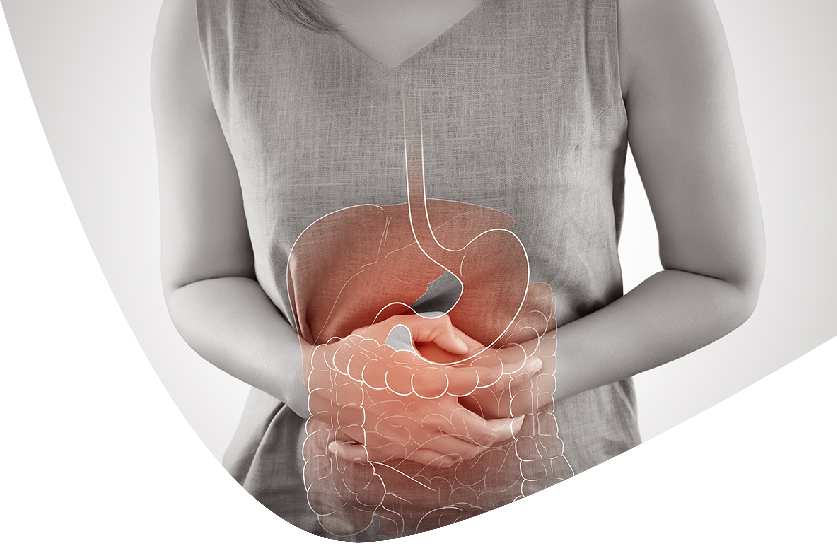
A 73-year-old female was confessed to our emergency clinic with stomach agony and looseness of the bowels. Processed tomography identified distension of the small digestive system. A palmar erythema, numerous oral ulcers, and desquamation of the fingers showed up after hospitalization. Little entrail endoscopic pictures showed different ulcers. We credited this case to disease with Yersinia pseudotuberculosis in view of the progressions in Y. pseudotuberculosis neutralizer titers over the span of the sickness.
Keyphrases: Small digestive system, Ulcers, Desquamation of the fingers, Yersinia pseudotuberculosis, Contamination, Kawasaki infectionPropels in clinical gadget improvement have empowered the assessment of sores in the small digestive tract that were already hard to survey. Small digestive system ulcers are delegate small-digestive tract sores, and their etiology can be idiopathic, irresistible, drug-instigated (ibuprofen and different NSAIDs), or avasculitis-related [1]. The two primary Yersinia species that are known to cause stomach side effects are Yersinia enterocolitica and Yersinia pseudotuberculosis. Purportedly, Y. pseudotuberculosis contamination is related with Kawasaki illness like sacred side effects [2, 3, 4, 5]. Notwithstanding, the hidden pathogenic instrument is indistinct. Moreover, it is expressed that there are no unmistakable contrasts in endoscopic pictures of patients with a disease by either microscopic organisms [6].
We report an instance of numerous small digestive system ulcers and desquamation of the fingers. After intensive examinations, the qualities of our case didn't meet the analytic standards for Kawasaki sickness. Furthermore, we felt that the etiology for this situation was probably going to be Y. pseudotuberculosis disease given its regular event in small digestive system ulcers and the adjustment of Y. pseudotuberculosis neutralizer titers in pair serum tests of the patient throughout the sickness. Both Kawasaki sickness and Y. pseudotuberculosis contamination are portrayed by a digestive system injury as the really established side effect. Be that as it may, as far as we could possibly know, no past reports have included endoscopic pictures of gastrointestinal sores brought about by Y. pseudotuberculosis disease. Subsequently, it is critical to extend the pathologic comprehension of these injuries by detailing the endoscopic pictures related with this problem.
A 73-year-old female was confessed to our medical clinic on May 2015. She introduced stomach torment and looseness of the bowels during 5 days, for which she was inspected and treated by an essential consideration doctor. As her side effects became worse, she was confessed to our clinic. She had a past filled with cholecystectomy 20 years sooner. She had no set of experiences of smoking or liquor misuse. She took no medicine. Moreover, she denied drinking great water.
On confirmation, her temperature was 37.7°C, her heartbeat was 90 pulsates/min, and her circulatory strain was 143/66 mm Hg. Her midsection was enlarged, however no other irregularity was seen upon actual assessment. Lab testing on affirmation showed an expanded C-receptive protein level. Distension of the small digestive system was additionally distinguished on registered tomography (CT).
The patient was at first overseen moderately. She got ceftriaxone 1 g/day by intravenous trickle implantation. Her C-responsive protein levels worked on briefly and afterward kept on expanding. A few different tests were performed however completely yielded adverse outcomes, for instance: bacterial societies of blood and stool, antinuclear antibodies, cytoplasmic enemy of neutrophil cytoplasmic neutralizer, myeloperoxidase ANCA, hostile to twofold abandoned DNA IgG immunizer, compound connected immunospot examine (T-SPOT®.TB), among others. A CT examine performed on hospitalization day 7 showed that the distension of the small digestive tract kept on declining. Stomach distension happened as an outcome of digestive system distension, and she in this manner went through ileus tube inclusion. On hospitalization day 12, her side effects started to move along. Fluoroscopy by means of the cylinder showed that there was no undeniable stenosis, yet there was stagnation of the differentiation medium. On hospitalization day 13, we played out a little gut endoscopy. We got endoscopic pictures of the impacted region that showed different injuries, including numerous ulcers running along the bearing of the minor pivot, map-like ulcers, and parts of upset mucous layer including the whole boundary of the ileum. On hospitalization day 20, we played out an esophagogastroduodenoscopy. We acquired endoscopic pictures of the mucous layer sores introducing as redness, disintegrations, and a ulcer reaching out from the duodenal angulus to the second piece of the duodenum . Be that as it may, no particular discoveries were gotten neurotically on hospitalization day 9, the patient introduced a palmar erythema, with the ensuing appearance of various oral ulcers on hospitalization day 11.Desquamation of the fingers happened on hospitalization day 13. We thought grown-up beginning Kawasaki infection or Y. pseudotuberculosis contamination as potential etiologies for the patient's condition in light of the presence of finger desquamation. A coronary corridor CT and an echocardiography were performed, yet these assessments uncovered no irregularities. Furthermore, there was no proof of coronary aneurysm. Consequently, in light of the clinical discoveries, our case didn't meet the demonstrative measures for Kawasaki sickness. Neutralizer titers of Y. pseudotuberculosis were estimated in pair serum tests showing a 80-crease increment on hospitalization day 11; these titers diminished from that point. Moderate clinical treatment was proceeded and the patient's side effects bit by bit gotten to the next level. She was released following 44 days of hospitalization.
We revealed an instance of different small digestive system ulcers related with desquamation of the fingers. As far as we could possibly know, our report is quick to portray such a show of Y. pseudotuberculosis contamination and give endoscopic pictures of the gastrointestinal sores. In spite of the fact that we couldn't disengage the bacterium, almost certainly, this was an instance of Y. pseudotuberculosis contamination for the accompanying reasons.
To start with, the patient introduced desquamation of the fingers. In view of the differential determination, we thought about Kawasaki sickness and Y. pseudotuberculosis contamination as the conceivable hidden etiologies. Desquamation of the fingers is profoundly unambiguous for Kawasaki illness and Y. pseudotuberculosis contamination. This side effect is seen in 96% of Kawasaki sickness cases and in 83% of Y. pseudotuberculosis contamination cases [7, 8].
Second, secluding the microorganisms in instances of Y is troublesome. pseudotuberculosis disease. We performed bacterial societies of stool, however the outcomes were negative. The presence of ileus blocked us from performing bacterial societies of biopsy tests right off the bat. Albeit a low-temperature culture is great, regardless of whether the way of life is kept up with at a low temperature and particular isolation culture medium is utilized, there is somewhere around a 40% probability of disconnecting these microbes [9]. Since we didn't think about Y. pseudotuberculosis contamination right on time during the illness course in the current case, we couldn't lead a low-temperature culture.
Third, subsequent to estimating Y. pseudotuberculosis immune response titers in pair serum tests of the patient, we noticed an ascent in the underlying levels with a resulting drop. We couldn't look at starting neutralizer titers given the postpone in laying out the essential conclusion and the titer change didn't arrive at 160-overlay increment. Notwithstanding, we emphatically think about that Y. pseudotuberculosis was the causal specialist in view of the 4-overlay immunizer titer ascend in just a single sort of the serum bunch. It appears to be far-fetched that such change in immunizer titers was owing to the presence of microscopic organisms other than Y. pseudotuberculosis.
We had the option to report the gastrointestinal injuries by endoscopy. The trademark endoscopic pictures of enteritis by Yersinia species contain terminal ileitis, dissipated mucosal ulcers with white overgrown appearance, ileocecal valve expanding, aphthous ulcers on the right digestive organ, and punctate unpredictable redness [10]. It has been accounted for that there is no reasonable contrast between the endoscopic qualities and conveyance of sores by Y. enterocolitica and Y. pseudotuberculosis [6]. Eminently, endoscopic pictures of cases like our own poor person been accounted for already. In a past report, the primary sore of enteritis by Yersinia was terminal ileitis [11, 12]. Matsumoto et al. [12] announced that these sores are changes of the singular lymph hubs and Peyer's fix. In the current case, the sores were broad in the duodenum and far reaching in the small digestive system. Moreover, the endoscopic pictures were likewise not the same as the delegate sores of Y. pseudotuberculosis contamination announced up to this point.
A few patients with Kawasaki sickness and Y. pseudotuberculosis contamination might give ileus, as in the current case [13, 14, 15, 16]. Miyake et al. [15] detailed that 7 of 310 patients with Kawasaki sickness created pseudo-check. For this situation, we noticed numerous ulcers running along the course of the minor hub, map-like ulcers, and mucous film disturbance including the whole periphery of the ileum. The instrument of such changes has been proposed to be mesenteric corridor vasculitis with entrail ischemia and related brokenness of the myenteric plexus [15, 17].
Neurotic assessment of the ischemic changes additionally shows vague incendiary cell invades comprising of lymphocytes and plasma cells [18]. The current case shows pathologic discoveries of ischemia. The endoscopic picture of the current case intently looks like ischemic enteritis. Hence, we consider that the endoscopic pictures of the digestive sores in the current case support the proposed component of ischemia prompting ileus auxiliary to Y. pseudotuberculosis contamination. The condition might deteriorate relying upon which region is impacted in connection with the mesentery. Besides, annular ulcers were blended in with map-like ulcers and villi decay.
Kawasaki illness is a fundamental vasculitis and a few instances of Y. pseudotuberculosis contamination show clinical circumstances like Kawasaki infection. Be that as it may, the etiology of the Kawasaki illness remains generally muddled. Past investigations have revealed a relationship between Kawasaki sickness and Y. pseudotuberculosis contamination [2,3,4,5]. In a past report, Y. pseudotuberculosis contamination was recognized in 9.3% of patients with Kawasaki illness [5]. Further, in certain reports, Kawasaki sickness symptomatic rules were met in 8.8-16.6% of cases with Y. pseudotuberculosis diseases [3, 19]. A few instances of Y. pseudotuberculosis contamination present with skin sores, and ileitis are remembered to foster optional to vasculitis set off by disease. We imagine that the endoscopic pictures acquired for our situation are quick to have caught these changes.
All in all, we noticed Y. pseudotuberculosis disease giving skin sores and ileus on endoscopic pictures. These endoscopic pictures proposed the presence of ischemic changes. Subsequently, our discoveries appear to demonstrate that a few instances of Y. pseudotuberculosis contamination, especially those giving skin sores and ileus, may foster vasculitis set off by the disease. Further, taking into account not just the qualities of Y is significant. pseudotuberculosis contamination, yet in addition those of Kawasaki illness, to comprehend these circumstances, which have covering introductions now and again. A few instances of Kawasaki illness include pseudo-deterrent, as in the current case. Moreover, it was hard to additional the comprehension of this condition, as it was viewed as a youth sickness as it were. We consider that even in Kawasaki illness with pseudo-impediment, there is plausible that the endoscopic pictures are like those saw for this situation. This report is important, as it delineates the endoscopic qualities of a Y. pseudotuberculosis contamination with foundational signs, which might empower us to develop the pathologic comprehension of this sickness.
1. Freeman HJ. Multifocal stenosing ulceration of the small digestive system. World J Gastroenterol. 2009;15:4883-4885. [PMC free article] [PubMed] [Google Scholar]
2. Konishi N, Baba K, Abe J, et al. An instance of Kawasaki illness with coronary corridor aneurysms recording Yersinia pseudotuberculosis disease. Acta Paediatr. 1997;86:661-664. [PubMed] [Google Scholar]
3. Baba K, Takeda N, Tanaka M. Instances of Yersinia pseudotuberculosis contamination having symptomatic rules of Kawasaki infection. Contrib Microbiol Immunol. 1991;12:292-296. [PubMed] [Google Scholar]
4. Sato K, Ouchi K, Komazawa M. Ampicillin versus fake treatment for Yersinia pseudotuberculosis disease in youngsters. Pediatr Contaminate Dis J. 1988;7:686-689. [PubMed] [Google Scholar]
5. Tahara M, Baba K, Waki K, et al. Investigation of Kawasaki sickness showing raised counter acting agent titres of Yersinia pseudotuberculosis. Acta Paediatr. 2006;95:1661-1664. [PubMed] [Google Scholar]
6. Okuyama Y, Shimizu S, Kohashi M, et al. Yersinia enterocolitis (in Japanese with English dynamic). Stomach Digestive system. 2008;43:1621-1628. [Google Scholar]
7. Wolff AE, Hansen KE, Zakowski L. Intense Kawasaki illness: not only for youngsters. J Gen Assistant Prescription. 2007;22:681-684. [PMC free article] [PubMed] [Google Scholar]
8. Satou K. Yersinia disease (in Japanese with English unique). Jpanes J Pediatr Prescription. 1983;21:1179-1183. [Google Scholar]
9. Matsumoto S, Hattori S. Yersinia disease (in Japanese with English unique). Jpn J Pediatr Drug. 2002;34:920-923. [Google Scholar]
10. Takahashi M, Ijyuu M. An instance of Yersinia enterocolitis (in Japanese with English dynamic). Gastroenterol Endosc. 2013;55:1866-1867. [Google Scholar]
11. Rutgeerts P, Geboes K, Ponette E, et al. Intense infective colitis brought about by endemic microorganisms in Western Europe: endoscopic highlights. Endoscopy. 1982;14:212-219. [PubMed] [Google Scholar]
12. Matsumoto T, Iida M, Matsui T, et al. Endoscopic discoveries in Yersinia enterocolitica enterocolitis. Gastrointest Endosc. 1990;36:583-587. [PubMed] [Google Scholar]
13. Zulian F, Falcini F, Zancan L, et al. Intense careful midsection as introducing appearance of Kawasaki sickness. J Pediatr. 2003;142:731-735. [PubMed] [Google Scholar]
14. Akikusa JD, Laxer RM, Friedman JN. Digestive pseudoobstruction in Kawasaki sickness. Pediatrics. 2004;113:504-506. [PubMed] [Google Scholar]
15. Miyake T, Kawamori J, Yoshida T, et al. Little gut pseudo-check in Kawasaki sickness. Pediatr Radiol. 1987;17:383-386. [PubMed] [Google Scholar]
16. Franken EA, Jr, Kleiman MB, Norins AL, et al. Gastrointestinal pseudo-check in mucocutaneous lymph-hub condition. Radiology. 1979;130:649-651. [PubMed] [Google Scholar]
17. Beiler HA, Schmidt KG, von Herbay A, et al. Ischemic little gut injuries for a situation of fragmented Kawasaki infection. J Pediatr Surg. 2001;36:648-650. [PubMed] [Google Scholar]
18. Okado Y, Takagi Y, et al. Ischemic enteritis (in Japanese with English theoretical). Stomach Digestive tract. 2016;51:1717-1719. [Google Scholar]
19. Sato K, Ouchi K, Taki M. Yersinia pseudotuberculosis contamination in kids, looking like Izumi fever and Kawasaki condition. Pediatr Taint Dis. 1983;2:123-126. [PubMed] [Google Scholar]
Azusa Kawasaki . Multiple small intestinal ulcers and finger desquamation. International Journal of Gastroenterology and Hepatology 2022.