Henry Ford Health System, 2799 West Grand Blvd. K-7, Detroit, MI 48202 (USA)
Henry Ford Health System, 2799 West Grand Blvd. K-7, Detroit, MI 48202 (USA)
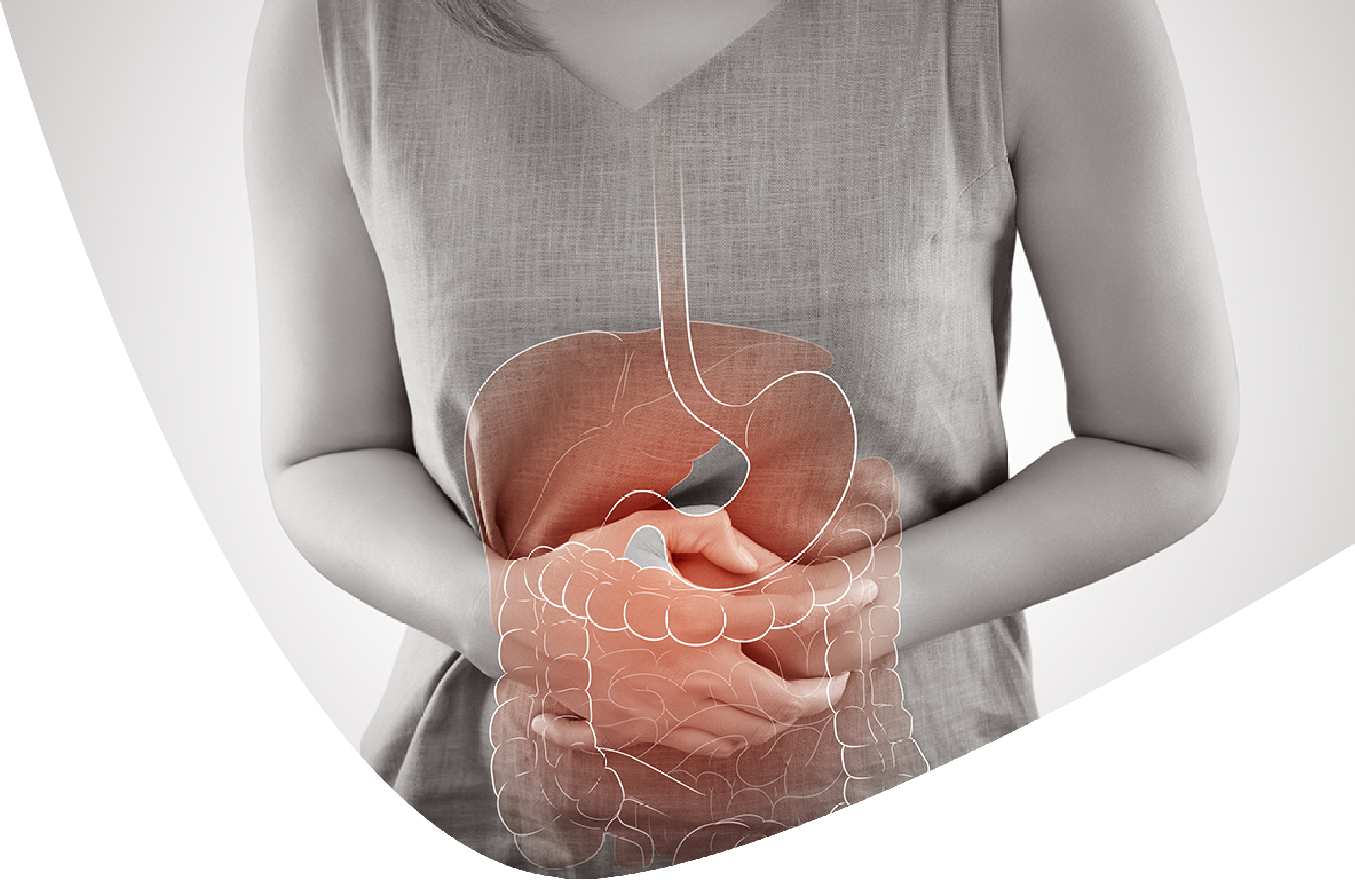
Intraductal papillary mucinous neoplasms (IPMNs) are mucin-creating papillary neoplasms of the pancreatic or biliary ductal framework that show variable cell atypia and cause ductal expansion. There are not many announced instances of IPMN emerging from the biliary tree in the writing. It has a higher inclination to go through dangerous change contrasted with IPMN emerging from the pancreatic conduit. A 80-year-old male went through cross-sectional tomography (CT) imaging of the mid-region for assessment of prostate adenocarcinoma, which uncovered a coincidental 2.3 × 2.7 cm delicate tissue mass focused at the porta hepatis with diffuse dilatation of the left intrahepatic biliary ductal framework and gentle conspicuousness of the right intrahepatic ductal situation. Endoscopic ultrasound showed 2 neighboring hilar masses including the normal hepatic pipe and the left hepatic conduit with projection of the tissue into the lumen of the channel and upstream ductal dilatation. Endoscopic retrograde cholangiopancreatography uncovered an enormous filling imperfection in the normal hepatic channel stretching out into the left hepatic pipe.
keyphrases: Intraductal papillary mucinous neoplasm, Bile pipe, Mucinous neoplasm
Intraductal papillary mucinous neoplasms (IPMNs) are mucin-delivering papillary neoplasms of the pancreatic or biliary ductal framework that display variable cell atypia and cause ductal enlargement. IPMN creates from epithelial cells and has harmful potential. It normally emerges from the pancreatic conduit [1]. IPMNs have been named primary conduit (MD) type or branch pipe (BD) type in view of the anatomic association of the pancreatic channel. Patients with inclusion of both the fundamental and branch conduits are alluded to as having blended type IPMN. The MD-IPMNs are histologically more forceful than BD-IPMNs and bound to hold onto a danger [2]. The male-to-female proportion for MD-IPMN has fluctuated in reports from 1.1 to 3: 1, and for BD-IPMN it has shifted from 0.7 to 1.8: 1 [3]. The gamble elements of IPMN incorporate cigarette smoking [4], diabetes, family background of pancreatic adenocarcinoma [5], Peutz-Jeghers condition [6], familial adenomatous polyposis disorder [7], or familial pancreatic carcinoma [8].
There are not many announced instances of IPMN emerging from the biliary tree in writing. It has a higher penchant to go through dangerous change contrasted with IPMN emerging from the pancreatic channel [9, 10]. Medical procedure is the therapy of decision because of the greater affinity of danger [11]. Our patient was found to have a coincidental biliary lot IPMN with high-grade dysplasia during the organizing of recently analyzed prostate adenocarcinoma.
A 80-year-old male was assessed for a raised prostate-explicit antigen level, and a biopsy of the prostate organ uncovered adenocarcinoma with a Gleason score 9. The patient achieved cross-sectional tomography (CT) imaging of the mid-region, which uncovered an accidental 2.3 × 2.7 cm delicate tissue mass focused at the porta hepatis with diffuse dilatation of the left intrahepatic biliary ductal framework and gentle unmistakable quality of the right intrahepatic ductal situation
On actual assessment, the head, eyes, ears, nose, and throat were ordinary. There was no jaundice. The mid-region was delicate, nontender, and nondistended, no masses or free liquid were found, and inside sounds were typical. Lab assessment uncovered ordinary liver compounds, lipase, and amylase. Sugar antigen (19-9) was 35.2 units/mL (ordinary worth 35 units/mL). The patient achieved endoscopic ultrasound and endoscopic retrograde cholangiopancreatography. Endoscopic ultrasound showed 2 contiguous hilar masses including the normal hepatic channel and left hepatic pipe with bulge of the tissue into the lumen of the conduit and upstream ductal dilatation with parenchymal decay. Endoscopic retrograde cholangiopancreatography uncovered an enormous filling deformity in the normal hepatic channel reaching out into the left hepatic pipe. A lot of cluster and delicate tissue with a fish-egg appearance was recovered. Two ponytail biliary stents were put into the left hepatic pipe upstream from the delicate tissue mass. The biopsy of the delicate tissue mass uncovered IPMN of the bile channel with diffuse high-grade dysplasia and intramucosal carcinoma.
The patient went through left hepatic lobectomy, extremist resection of the normal hepatic channel with Roux-en-Y hepaticojejunostomy to the right hepatic conduit. Histopathological assessment of the resected example uncovered IPMN with diffuse high-grade dysplasia. The careful edges and lymph hubs were negative for carcinoma.
The postoperative course was convoluted by intraabdominal abscesses and pneumonia, requiring intravenous anti-infection agents and CT-directed seepage. The patient was readmitted fourteen days after release with demolishing hypoxia and lung penetrates. He was found to have eosinophilic pneumonitis auxiliary to the anti-microbials daptomycin and meropenem that settled with stopping of the prescriptions.
A subsequent CT sweep of the mid-region 2 months after the medical procedure was negative for any masses. The patient feels good and stays asymptomatic a year after the medical procedure.
Intraductal papillary mucinous neoplasms (IPMNs) are intraductal, mucin-creating growths emerging from epithelial cells and have dangerous potential. IPMN for the most part emerges from the pancreatic pipe. In the writing, there are not many announced instances of IPMN emerging from the biliary tree [9, 11, 12, 13, 14].
IPMN of the bile pipe is typically connected with more elevated levels of starch antigen 19-9 and carcinoembryonic antigen. It has a higher inclination to go through threatening change contrasted with IPMN emerging from the pancreatic conduit [10, 11]
It creates a lot of mucin, prompting mass impact and unsettling influences in the bile stream. It is related with bile pipe dilatation and seldom shows intrusion of encompassing edges. The most well-known type of show is a harmless neoplasm. In uncommon cases, bile channel dilatation is the main finding. It can prompt blister development or papillary multiplication and may have fluctuating levels of cell atypia. The threatening possible ascents with expanding size (>3 cm) [14, 15]. Various hereditary transformations have been portrayed [16].
Most IPMNs of the biliary tree are harmless and painless; in any case, because of expanding size and threatening potential, careful resection is the treatment of decision. The careful treatment depends on the area of the IPMN. Different careful medicines incorporate resection of a liver curve, hepaticojejunostomy, pancreaticoduodenectomy, and bile conduit resection [10, 15]. The patients with harmless neoplasms have a decent long haul guess and endurance.
There is no known relationship between prostate malignant growth and biliary pipe IPMN in the writing. The conclusion of IPMN in our patient was coincidental and didn't appear to be connected with the patient's basic prostate adenocarcinoma.
1. Adsay V, Mino-Kenudson M, Furukawa T, Basturk O, Zamboni G, Marchegiani G, et al. Pathologic assessment and announcing of intraductal papillary mucinous neoplasms of the pancreas and other tumoral intraepithelial neoplasms of pancreatobiliary plot: suggestions of Verona agreement meeting. Ann Surg. 2016;263:162-177. [PMC free article] [PubMed] [Google Scholar]
2. Serikawa M, Sasaki T, Fujimoto Y, Kuwahara K, Chayama K. The executives of intraductal papillary-mucinous neoplasm of the pancreas: treatment technique in view of morphologic characterization. J Clin Gastroenterol. 2006;40:856-862. [PubMed] [Google Scholar]
3. Ingkakul T, Warshaw AL, Fernandez-Del Castillo C. The study of disease transmission of intraductal papillary mucinous neoplasms of the pancreas: sex contrasts between 3 geographic locales. Pancreas. 2011;40:779-780. [PubMed] [Google Scholar]
4. Fukushima N, Mukai K. Pancreatic neoplasms with plentiful bodily fluid creation: accentuation on intraductal papillary-mucinous growths and mucinous cystic cancers. Adv Anat Pathol. 1999;6:65-77. [PubMed] [Google Scholar]
5. Capurso G, Boccia S, Salvia R, Del Chiaro M, Frulloni L, Arcidiacono PG, et al. Risk factors for intraductal papillary mucinous neoplasm (IPMN) of the pancreas: a multicentre case-control study. Am J Gastroenterol. 2013;108:1003-1009. [PubMed] [Google Scholar]
6. Sato N, Rosty C, Jansen M, Fukushima N, Ueki T, Yeo CJ, et al. STK11/LKB1 Peutz-Jeghers quality inactivation in intraductal papillary-mucinous neoplasms of the pancreas. Am J Pathol. 2001;159:2017-2022. [PMC free article] [PubMed] [Google Scholar]
7. Maire F, Hammel P, Terris B, Olschwang S, O'Toole D, Sauvanet A, et al. Intraductal papillary and mucinous pancreatic growth: a new extracolonic cancer in familial adenomatous polyposis. Stomach. 2002;51:446-449. [PMC free article] [PubMed] [Google Scholar]
8. Poley JW, Kluijt I, Gouma DJ, Harinck F, Wagner A, Aalfs C, et al. The yield of first-time endoscopic ultrasonography in screening people at a high gamble of creating pancreatic disease. Am J Gastroenterol. 2009;104:2175-2181. [PubMed] [Google Scholar]
9. Subhash R, Valiyaveettil IA, Natesh B, Raji L. Biliary parcel intraductal papillary mucinous neoplasm: a short report and survey of writing. Indian J Pathol Microbiol. 2014;57:588-590. [PubMed] [Google Scholar]
10. Minagawa N, Sato N, Mori Y, Tamura T, Higure A, Yamaguchi K. A correlation between intraductal papillary neoplasms of the biliary lot (BT-IPMNs) and intraductal papillary mucinous neoplasms of the pancreas (P-IPMNs) uncovers unmistakable clinical signs and results. Eur J Surg Oncol. 2013;39:554-558. [PubMed] [Google Scholar]
11. Barton JG, Barrett DA, Maricevich Mama, Schnelldorfer T, Wood CM, Smyrk TC, et al. Intraductal papillary mucinous neoplasm of the biliary lot: a genuine sickness? HPB (Oxford) 2009;11:684-691. [PMC free article] [PubMed] [Google Scholar]
12. Mo A, Whelp G, Spolverato G, Pawlik TM. Intraductal papillary mucinous neoplasm of the liver: GI picture. J Gastrointest Surg. 2015;19:792-794. [PubMed] [Google Scholar]
13. Wang X, Cai YQ, Chen YH, Liu XB. Biliary parcel intraductal papillary mucinous neoplasm: report of 19 cases. World J Gastroenterol. 2015;21:4261-4267. [PMC free article] [PubMed] [Google Scholar]
14. Rocha FG, Lee H, Katabi N, DeMatteo RP, Fong Y, D'Angelica MI, et al. Intraductal papillary neoplasm of the bile pipe: a biliary identical to intraductal papillary mucinous neoplasm of the pancreas? Hepatology. 2012;56:1352-1360. [PubMed] [Google Scholar]
15. Jung G, Park KM, Lee SS, Yu E, Hong SM, Kim J. Long haul clinical result of the carefully resected intraductal papillary neoplasm of the bile channel. J Hepatol. 2012;57:787-793. [PubMed] [Google Scholar]
16. Sclabas GM, Barton JG, Smyrk TC, Barrett DA, Khan S, Kendrick ML, et al. Recurrence of subtypes of biliary intraductal papillary mucinous neoplasm and their MUC1, MUC2, and DPC4 articulation designs contrast from pancreatic intraductal papillary mucinous neoplasm. J Am Coll Surg. 2012;214:27-32. [PubMed] [Google Scholar]
Ravish Parekh . Rare Case of Biliary Duct Intraductal Papillary Mucinous Neoplasm in a Prostate Adenocarcinoma Patient. International Journal of Gastroenterology and Hepatology 2022.